- home
- Parrots
- Diseases in parrots
- Eye diseases in parrots
Modern veterinary medicine has become concerned with the treatment of eye diseases in birds relatively recently.
The differences in the features, functions and structure of the eyes between birds and animals are enormous, and therefore diagnostic and treatment methods used for mammals cannot be applied to birds.
The most important research in the field of ornitho-ophthalmology is carried out at the Institute of Avian Diseases in Munich.
Dr. Rüdtger Korbel has created special methods for studying birds and their treatment, which, quite possibly, will soon begin to be put into practice throughout the world.
Symptoms
The symptoms of the disease are varied, and it is impossible for the owner to make a diagnosis without the appropriate education. Let's look at the most common diseases found in parrots.
Conjunctivitis and inflammation of the eyelid - develops as a result of hypothermia and poor hygiene. Manifestations include swelling of the eyelids, discharge of fluid from the eyes, and redness.
Third eyelid prolapse is identified by thickening, swelling and redness. Develops as a result of ingress of a foreign object.
Another frequently diagnosed pathology is cystosis, which a veterinarian determines by swelling of the lower eyelids. Cystosis is often confused with other ailments, and only an experienced ornithologist can make a correct diagnosis.
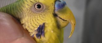
Xerophthalmia - occurs due to vitamin A deficiency, as well as from a chemical burn. The parrot's vision deteriorates, photophobia develops, the cornea dries out, becomes hardened, and color changes.
What eye diseases are there in parrots?
The eyes are one of the first to respond to infection in the body.
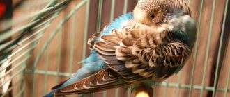
- Often the eyes of a budgie suffer because of the owners themselves, who either smoke in the presence of the pet or create dust. Moreover, the dust can be not only due to the dry air in the room, but due to the fact that very fine sawdust is used as bedding. As a result, the eyes begin to water and the skin around them turns red. Although the disease is non-infectious, it should not be left to chance. After all, sometimes the inflammatory process from the eyes “transfers” to the respiratory organs. And this can lead to death.
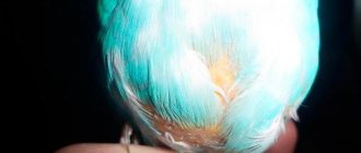
- If we talk about non-contagious eye diseases of parrots, then it is imperative to know about hypovitaminosis. A lack of vitamin A affects not only visual acuity, but also the condition of the parrot's eyes themselves. If an animal is deficient in this vitamin, the mucous membranes suffer. They dry out. The bird's organ of vision becomes dull and practically does not shine. Your pet will try to blink more often, tears will flow in order to somehow “wet” the parrot’s eyes.
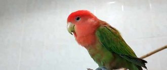
The parrot has a bright look
- You should not keep your feathered pet’s cage in drafts or in a very cold room. This will lead to eye diseases. The little beads on your parrot's face will become inflamed very quickly. Conjunctivitis will develop. But it is worth understanding that it can be not only the main disease, but also an important symptom of something else. It is important not to miss this, otherwise the infection will spread throughout the body, and the consequences will be disastrous.
- Eye injury. The bird could accidentally stumble upon a sharp twig, scratch with its paw and catch the cornea with its claw. Or if there is another pet, then a fight could arise between them. And one will peck the other in the eye. There are actually a lot of opportunities to get injured. You yourself are unlikely to see a scratch on the cornea. It is better to take it to an ornithologist so that he can carefully examine one eye.
- Eye diseases of parrots can be of contagious etiology (bacterial, viral, fungal, even parasitic). To clarify the diagnosis, be sure to visit a qualified ornithologist veterinarian. Do not try to guess what your pet is sick with on your own. Moreover, do not even try to prescribe and use medications, drops, ointments. Only a doctor can prescribe medications and procedures.
Treatment
Any drugs are administered in the form of drops. The selection of drops should only be performed by an experienced veterinarian.
Unlike mammals and humans, ointments cannot be used - they provoke gluing of the plumage, which in turn provokes the development of a secondary infection.
In addition to instilling drops, they resort to rinsing. If infection is present, antibiotics are prescribed.
Foreign bodies often get in, which the doctor removes with special tweezers under anesthesia.
For a number of ailments, a multivitamin complex that includes vitamin A is prescribed. The drugs are given in the form of drops or injections.
Panophthalmitis, provoked by injuries to the eyeball, as well as sinusitis in an advanced stage, has symptoms such as protrusion of the eyeball, the lens becomes cloudy, and the cornea is destroyed. There is a high risk of complete blindness.
The most severe and irreversible pathology, blindness, often occurs for no apparent reason at first glance.
In fact, blindness is caused by age-related changes, oncology, and poor indoor lighting.
Molluscum contagiosum in children
Molluscum contagiosum is more common in children than in adults. Children up to one year old practically do not get sick with molluscum contagiosum. This is due to the fact that in the first year of life the child’s circle of contacts is small: the child moves in a limited space, often specially prepared for him and under the strict supervision of adults, without trying to come into contact with other children. But as soon as a child begins to actively communicate and independently explore the world, the threat of becoming infected with molluscum contagiosum increases sharply.
The peak incidence of molluscum contagiosum in children occurs between the ages of 2 and 6 years. Immunity at this age is still weak. Children become infected through toys or dirty hands. The virus penetrates the skin in the place where the integrity of the skin is damaged - through wounds, abrasions, cracks. Children's skin is delicate and sensitive, and the activity of a preschool child is great. As a result, numerous microtraumas occur, opening the way for infection. Cases of infection with molluscum contagiosum while swimming in the pool have also been described.
From 6 to 10 years, the incidence of molluscum contagiosum decreases. Instilling household hygiene skills is of great importance. The sooner your child starts taking care of clean hands, the better.
Possible complications
The main complication after eye diseases is deterioration or complete loss of vision.
If you consult a specialist in a timely manner, there is a chance to avoid them and preserve your pet’s ability to see.
Call a veterinarian to your home 24 hours a day
The clinic’s specialists urge you not to delay seeking help if you suspect eye pathology.
The veterinary center staff will come to your home at any time of the day, make a diagnosis, provide first aid and select therapy.
- You can choose the appropriate day for the ornithologist’s visit.
- The pet will remain at home in a comfortable environment and will experience less stress from meeting with the veterinarian.
- The cost of home care is no higher than in a veterinary center; there are different payment methods.
- There is an emergency team that goes to the site at any time of the day.
- Most therapeutic and diagnostic procedures are performed at home.
To invite a doctor and get advice on our services, call the numbers listed on the website.
General services
| Prices for services in our clinic | In the clinic and at home |
| Ornithologist visiting your home | from 500 |
| Clinical examination, preliminary diagnosis, consultation | from 500 |
| Telephone consultation | for free |
| Therapy | from 150 |
| Surgery | from 150 |
| Ambulance at home (within an hour) | from 1000 |
Therapy
| Prices for services in our clinic | In the clinic and at home |
| Subcutaneous administration of medication to birds | from 200 |
| Intramuscular administration of medication to birds | from 150 |
| Intravenous administration of the drug to birds | from 500 |
| Bird dropper | from 1000 |
| Administration of the drug intraperitoneally, intraosseously | from 800 |
| Nerve receptor blockade in birds | from 500 |
| Tube feeding of birds | from 300 |
| Cleaning the horny cover | from 400 |
| Resuscitation treatment of birds | from 1500 |
| Infusion therapy | from 250 |
| Intravenous catheter placement | 500 |
| Removing the IV catheter | 500 |
| Removing the Marking Ring | from 200-1000 |
| Taking blood for laboratory tests | from 350 |
| Obstetrics in birds | 1 hour from 500 |
| Washing the crop | from 500 |
| Trimming: | |
| Beak | 500 |
| Claws | 500 |
| Krylyev | 500-1000 |
| Tail | 500-1000 |
Surgery and Traumatology
| Prices for services in our clinic | In the clinic and at home |
| Surgical treatment of wounds | from 200-1000 |
| Stitching | from 300-1500 |
| Application of a splint | from 300 |
| Opening abscesses, hematomas | from 300 |
| Removal of tumors | from 1000 |
| Goiter surgery | from 1500 |
| Cloaca surgery | from 1500 |
| Removal of foreign bodies | from 1500 |
| Eye amputation | from 2000 |
| Pelvic limb amputation | from 2500 |
| Osteosynthesis | from 10000-30000 |
| Surgery for goiter rupture | from 1500 |
| Egg extraction | from 500 |
| Anesthesia for birds | from 1500-3500 |
| Puncture of air sacs | from 1000 |
| Abdominal wall puncture | from 1000 |
Anesthesiology for birds
| Prices for services in our clinic | In the clinic and at home |
| Anesthesia | from 500 |
Ophthalmology
| Prices for services in our clinic | In the clinic and at home |
| Eyelid surgery for inversion, eversion | from 2000 |
| Exenteration of the eyeball | from 3000 |
Laboratory research
| Prices for services in our clinic | In the clinic and at home |
| Taking tests | 500 |
| Clinical blood test | 1000 |
| General clinical blood test | 1000 |
| Blood chemistry | 2000 |
| Microscopy of blood parasites | 700 |
| Smear microscopy | 700 |
| Examination of droppings for protozoa | 1000 |
| Examination of droppings for helminths | 700 |
| Determination of gender | 2000 |
| Histological examination | 2500 |
| Examination of droppings for helminths | 700 |
| Infectious anemia of chickens | 1500 |
| Test for infectious encephalomyelitis | 1500 |
| Analysis for infectious bronchitis of birds | 1500 |
| Analysis for infectious rhinotracheitis in birds | 1500 |
| Salmonellosis test | 1500 |
| Testing for trichomoniasis | 1500 |
| Tuberculosis test | 1500 |
| Examination of droppings for protozoa | 1000 |
| Analysis for psittacosis | 1500 |
| Analysis for pasteurellosis | 1500 |
| Avian influenza test | 1500 |
| Analysis for dysbacteriosis | 2000 |
| Analysis for avian mycoplasmosis | 1500 |
| Newcastle disease test | 1500 |
| Analysis for avian leukemia | 1500 |
| Test for Marek's disease | 1500 |
| Analysis for avian adenovirus | 1500 |
| Analysis for avian circovirus | 2000 |
| Analysis for yeast-like fungi | 1700 |
| Comprehensive analysis for anthropozoonoses | 4000 |
| General bacteriological analysis | 3000 |
| Gumboro disease | 1500 |
| Avian reovirus | 1500 |
What is otitis media
Otitis is the general name for inflammatory processes in the ear area.
Inflammation can be acute or chronic, affecting various parts of the ear. If the inflammation is localized in the auricle and ear canal to the border of the eardrum, this is otitis externa; inflammation in the tympanic cavity is otitis media; if the area of the cochlea is affected, the inner part of the ear is internal otitis or labyrinthitis.
These pathologies are extremely painful, accompanied by fever, hearing impairment, and discharge from the external meatus. In addition, without treatment, otitis media can lead to serious complications - hearing loss or complete deafness, paresis in the area of the facial nerve, damage to the bones or brain.
What antibiotics are effective for otitis media?
“Systemic antibacterial therapy is indicated in all cases of moderate and severe acute otitis media,” says otolaryngologist Svetlana Kovaleva, “as well as in patients with immunodeficiency conditions.
If otitis is mild (no pronounced symptoms of intoxication, pain, hyperthermia up to 38 ° C), you can refrain from prescribing antibiotics. However, if there is no positive dynamics within 48 hours, antibiotic therapy should be resorted to. For otitis, broad-spectrum antibiotics are prescribed that are effective against typical pathogens: Streptococcus pneumoniae, Haemophilus influenzae, Moraxella catarrhalis, Streptococcus pyogenes, Staphylococcus aureus.
The drug of choice is Amoxicillin.
Alternative drugs for allergies to β-lactams are modern macrolides (Josamycin, Azithromycin, Clarithromycin). In case of ineffectiveness, as well as in patients who have received antibiotics for a month, for patients over 60 years of age, it is advisable to prescribe a complex - amoxicillin + clavulanic acid. Alternative drugs are II-III generation cephalosporins (Cefuroxime axetil, Ceftibuten) or fluoroquinolones (Levofloxacin, Moxifloxacin).
For mild to moderate cases, oral antibiotics are indicated. In severe and complicated cases of otitis, begin with intravenous or intramuscular administration of the drug, and then continue treatment orally.
The duration of antibacterial therapy is 7–10 days. For complicated otitis – 14 days or more.
You should not use antibiotics on your own; you should consult an otolaryngologist. Otitis media can be caused by fungal flora or herpes infection. The use of antibiotics in this case can worsen the course of the disease.
Is stye contagious?
It is not dangerous for others, but only if basic hygiene rules are followed. The risk of infection usually occurs in young children. Due to their age, they do not yet understand the importance of hygiene and therefore often suffer from bacterial infections. Infection is also possible when the patient uses cosmetics. But in general, isolation of an adult or child suffering from hordeolum is not required.
The causative agents of the disease are found in pus located in the barley sac. There are no bacteria on the surface. But there is usually little pus inside, so even a burst stye is not dangerous. In 80% of cases, the abscess breaks out at night when the patient is sleeping. If purulent masses end up anywhere, it is on a person’s face, his pillow, or bed linen. The infection does not spread further.
Types and stages of development of hordeolum
Depending on the location of the lesion, it can be of the following types:
- External stye
. More common according to statistics. The infectious-inflammatory process is localized at the edge of the eyelid, since the infection settles in the sebaceous gland of Zeiss or in the ciliary bulb. - Internal stye on the eye
. It has another name - meibomite. It is caused by the penetration of pathogenic microorganisms into the passage of the meibomian glands located on the back side of the eyelid margin.
By type they distinguish:
- Hot barley
. It is characterized by the classic development of the disease; infection is localized in the ciliary pocket - the bulb or Zeiss gland. With therapy it goes away in about a week. - Cold stye (chalazion, meibomian cyst)
. The inflammatory process affects the meibomian glands. It develops very slowly. Recovery takes 1–2 months. If there is a very large stye, surgical removal is possible.
Inflammation goes through the following stages:
- Formation of a purulent core
. A small reddish swelling causes discomfort when moving the eyelids. - Formation of an abscess
. The patient’s condition at this stage can only be alleviated by medications. - Breakthrough of pus.
It does not indicate recovery, but the patient feels much better. During this period, you need to keep your eyes clean and use prescribed medications and ointments.
Diagnostics
The diagnosis can be suspected based on typical complaints, but the doctor will ask in detail where and how the ear hurts, press on the tragus, pull the earlobe down to determine whether there is pain. In addition, the otorhinolaryngologist will examine the ear using instruments and lighting to specifically examine the ear canal, eardrum, and determine whether there is pus or perforation in it. To determine sensitivity to antibiotics, flora culture is performed. The doctor may also prescribe:
- blood tests (general, biochemistry) to determine the nature of inflammation;
- X-ray of the paranasal sinuses, if a connection with sinusitis is suspected;
- X-ray of the temporal bone in chronic otitis media.
All this data is needed in order to determine treatment tactics, the need for antibiotics, surgical interventions (membrane perforation or other interventions).
Treating Watery Eyes: What You Can Do
With age, the quality of tear fluid deteriorates. Our tear fluid contains more than just a water component. Our body produces a mixture to moisturize the eyes. If your eyes don't get the moisture they need, they may compensate by producing tears, resulting in watery eyes.
If this is a chronic condition, your doctor can recommend possible treatments. Action needs to be taken.
The cause of watery eyes can be easily treated, so do not delay treatment. Even if surgery is required, it is usually an outpatient procedure. Why not do this at an early stage?
Prevention of otitis in adults at home
To prevent otitis, it is necessary to avoid hypothermia, wash your hands after going outside, irrigate the nasal mucosa with sea water after visiting places with large crowds of people, exercise, exercise, and eat fresh fruits, vegetables, and dairy products every day.
If it so happens that you get sick and a runny nose begins to bother you, then you need to blow your nose extremely carefully, while freeing only one nostril, otherwise nasal discharge can get through the auditory tube into the ear and provoke otitis media.
Proper ear hygiene is essential. It is not recommended to use cotton swabs - they can introduce a bacterial or fungal infection into the ear. For ear hygiene, use drops consisting of a combination of surfactants (Allantoin, Benzetoin chloride) that clean, moisturize and protect the skin of the external auditory canal.
Preventive measures
Prevention includes following the recommendations:
- Strengthening immune forces
. This should always be done, not only when there is a threat of hordeolum or in the off-season. It is recommended to take vitamins, give preference to fresh vegetables and fruits, move more, be in the fresh air, and follow a daily routine. - Timely elimination of ophthalmological disorders
. If you are prone to eye diseases, you should regularly visit an ophthalmologist. Self-diagnosis and self-medication usually contribute to the transition of pathology to a chronic form. - Compliance with hygiene standards
. You need to have your own towel, wash your hands with soap, change bed linen every 1–2 weeks, and not use other people’s cosmetics. - Developing the habit of not touching your eyes
. This should not be done even in a normal state, when a person is not bothered by any problems. This rule also applies to the face - you should not touch it during the day with unwashed hands.
Simple preventive measures will help avoid inflammation and keep your eyes healthy.