The eyes of a parrot are a very important organ that helps birds easily navigate the space around them.
At the same time, it is worth immediately focusing your attention on the fact that the vision of birds is structured somewhat differently than that of humans. Surely you have noticed more than once that your ward easily navigates during his flight, skillfully maneuvering between moving people and at the same time choosing a place to land.
In addition, while eating delicacies offered by people, parrots constantly inspect the entire space around them. After all, only thanks to their observation skills, birds living in the wild can avoid meeting a predator sneaking up on them and fly away.
However, like other living creatures, the eyes of feathered pets can suffer from a number of threats, some of which we will list in the following article.
Inflammatory processes
It is not for nothing that we have placed various inflammatory processes in the first place on the list of circumstances dangerous to a pet’s eyes. After all, they can be caused by completely different factors, which every bird breeder should be aware of.
If there is at least one smoker in your family and he does not bother to get ready and go outside or onto the balcony to smoke, then most likely it is very smoky in your apartment.
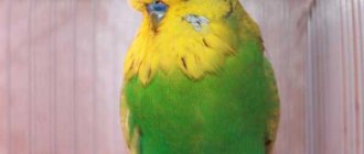
Let’s not talk now about the dangers of smoking for people, because we are not talking about them, but about small and defenseless birds, who perceive tobacco smoke much more painfully. It is this that often becomes the reason that your feathered charges, for no apparent reason, begin to get red and inflamed eyes.
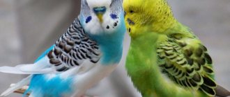
Treatment and prevention of eye diseases in budgies
Firstly, you should not self-medicate. While it is still possible to somehow treat non-contagious conjunctivitis yourself, infectious types of diseases can only be accurately diagnosed by a doctor.
Secondly, provide the bird with normal living conditions. The cage should not be placed where there are drafts. The bird should also be protected from smoke and other strong odors. Therefore, the kitchen is a completely unsuitable place for a budgie to live. In a room suitable for it, clean it more often so that the space does not become dusty.
Thirdly, if you decide to get another pet, then the newcomer needs to be kept in quarantine for a month and only then can you put it in a cage with another bird. This way you will avoid over-infection of the parrots.
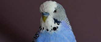
And, of course, be attentive to your birds. Monitor their condition, appearance, unusual behavior, and ensure that the budgerigar's eyes remain shiny black beads.
After all, it is always easier to treat a disease at the very beginning than in an advanced stage.
Foreign body
Surely you yourself have often gotten some kind of midge or speck into your eye. All of them evoke a persistent desire to remove them from the eye. Without thinking about the consequences of the fact that you begin to strongly rub the eye that is bothering you.
When a parrot encounters a similar phenomenon, it, just like other living beings, tries at all costs to get rid of the speck that has gotten into its eye.
Observing this from the side, you can see how the bird is intensely itching against the twigs or trying to get rid of irritation in some other way accessible to birds. Moreover, this often leads to the fact that the eye turns red and an inflammatory process begins in it, as in the case of colds. It often happens that birds' third eyelids swell and fall out.
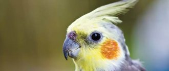
Xerophthalmia
The disease is possible due to a lack of vitamin A in the body or a chemical burn to the eye. Translated from Greek, xerophthalmia means “dry eye.”
Symptoms:
- Cloudiness, drying of the cornea;
- Hardening, desquamation, discoloration of the cornea;
- Deterioration of vision;
- Fear of light;
- Complete or partial absence of pupil constriction in bright light.
Treatment consists of a multivitamin complex with the obligatory presence of vitamin A in the form of drops or injections.
Cystosis
To some extent, this disease is similar to conjunctivitis. They can be distinguished by the area of inflammation. If conjunctivitis affects the entire organ of vision, then with cystosis the inflammation affects exclusively the lower eyelid.
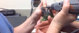
If a parrot's eye is closed, and the lower eyelid is swollen and a purulent accumulation is visible inside it, this is cystosis. The parrot needs urgent professional treatment. It is worth noting that the problem of suppuration in the eyelid with cystosis can only be solved by surgical intervention.
Risk factors
Some pathological conditions and unfavorable factors can lead to the development of functional disorders of the visual analyzer:
- ametropia - deviation of the refractive power of the eye from age-related values;
- strabismus;
- cerebral palsy;
- mental development disorder;
- III and IV degree of prematurity of the child;
- underweight newborn;
- the presence of ophthalmological diseases in close relatives (amblyopia, strabismus, lens opacity).
The presence of bad habits in pregnant women (smoking and drinking alcohol) increases the risk of developing visual system dysfunction several times.
Blindness
Without obvious causes, progressive blindness can be the result of a tumor in the brain, damage to the optic nerve, old age, or poor lighting. The parrot bumps into objects, cannot find food, and the pupil does not react to bright light. Treatment is ineffective.
Eye diseases can be not only a sign of a problem with them, but also a consequence of infections or disturbances in the functioning of other organs. Therefore, you should not start self-medication. It is better to consult a specialist to identify the true causes of the ailment and receive the correct treatment.
Determination of the angle of strabismus
When amblyopia develops against the background of strabismus, the doctor determines the angle of strabismus according to Hirschberg. To do this, the child is asked to look at the ophthalmoscope, after which the doctor records the light reflections. Normally, the glare is projected in the middle of the pupil, but with strabismus - at the edge of the pupil, within or beyond it, which determines the angle of deviation.
Determining the nature of visual fixation is the most important diagnostic procedure for any form of amblyopia, since the treatment regimen depends on visual fixation.
Get a complete vision examination at the Lege Artis Eye Clinic
It's time to correct your vision!
Make an appointment by phone:
8(804) 333-02-14 Free call
Floaters before eyes
Atherosclerosis
Diabetes
66848 December 14
IMPORTANT!
The information in this section cannot be used for self-diagnosis and self-treatment.
In case of pain or other exacerbation of the disease, diagnostic tests should be prescribed only by the attending physician. To make a diagnosis and properly prescribe treatment, you should contact your doctor. Flashing of flies before the eyes: reasons for their appearance, what diseases they occur with, diagnosis and treatment methods.
Definition
The flickering of flies before the eyes is a subjective sensation, when describing it, people imply the presence in the field of vision of permanent or transient additional inclusions of various shapes and sizes.
The organ of vision is one of the most important sense organs providing orientation in space. The visual analyzer consists of the organ of vision (eye), which perceives and interprets the image into an electrical signal, the optic nerve, through which the signal enters the subcortical centers, and the cerebral cortex, where the signal is analyzed. The eye consists of the eyeball and accessory organs, and the eyeball is made up of three membranes: outer (cornea and sclera), middle (iris and choroid), and inner (retina). Behind the iris is the crystalline lens, a light-refracting lens. The space between the lens and the retina is filled with vitreous humor.
The vitreous body is a gel-like structure enclosed in a dense membrane. The gel contains water, hyaluronic acid, collagen fibers, proteins and other substances.
If the ratio of certain components of the vitreous body is disturbed, dense inclusions can form, perceived by a person as “spots”.
Types of flies before the eyes
“Flies” are dots, zigzags, worms, dust, dashes, strokes, scratches, cobwebs, commas, etc.
Floaters before the eyes, arising as a result of destructive processes in the vitreous, have clear descriptions and do not disappear from view:
- rings with an outer and inner rim or a dot inside, located in the field of view in large numbers, may indicate granular destruction of the vitreous body;
- threads that look like short or long strips with circles inside, of varying transparency, may indicate filamentous destruction of the vitreous body. The interweaving of threads indicates more severe destruction.
Destructive processes in the vitreous body Also, with the destruction of the vitreous body, spots and clots may appear, similar to “plastic film” or flakes, which greatly cloud the image.
When cholesterol crystals are included in the vitreous gel, shiny “snowflakes” appear in the field of view.
Any inclusions can move freely throughout the entire field of view, move within a limited range, or be stationary.
Possible causes of flies flashing before the eyes
The reasons for flies flashing before the eyes are quite varied. Floaters can be a sign of temporary functional disorders, for example, when lifting weights, overwork, or a sudden change in posture. However, inclusions in the visual field appear in some diseases of the eye structures - the vitreous body, choroid, retina, and also arise as a result of diseases of other organs and systems that affect the functioning of the visual analyzer.
What diseases cause floaters to appear before the eyes?
Destruction of the vitreous body
is the reason for the constant presence of additional inclusions in the field of view. It occurs due to a decrease in the ratio of fluid and other components of the vitreous gel during natural aging or after eye injury, eye surgery and other reasons. The characteristics of inclusions are described in the previous section.
The prognosis for vision is favorable, but the quality of life may suffer and the ability to work may decrease.
Retinal and/or vitreous detachment
, in addition to floaters, flashes and sparks, is characterized by a sharp deterioration in visual acuity and blurred vision. Without immediate medical attention, it can lead to blindness. The risk of detachment increases in people suffering from a high degree of myopia.
For vertebrobasilar insufficiency
blood flow through the vertebral arteries is disrupted, which affects the blood supply to certain areas of the brain, including those responsible for visual function. Clinically manifests itself as floaters, darkening before the eyes, short-term loss of vision.
The most common cause of acquired vertebrobasilar insufficiency is osteochondrosis of the cervical spine.
Choroiditis (inflammation of the choroid), retinitis (inflammation of the retina), chorioretinitis (inflammation of both structures)
can also cause the appearance of floaters. In addition, the patient experiences flashes of light (photopsia), visual acuity decreases, and vision in the dark is impaired. The disease of an infectious nature in advanced cases leads to blindness.
Retinopathy
(hypertensive, atherosclerotic, diabetic) occur when blood vessels are damaged by diseases of the same name. Retinopathy is characterized by transient visual impairment in the form of decreased visual acuity, floaters before the eyes, and the presence of large spots in the field of vision (scotomas). As the underlying disease is treated, the intensity of the symptoms decreases, and as it progresses, it can result in loss of vision.
Transient flashing of flies before the eyes, a feeling of a “noisy” picture can occur with low blood pressure.
Pressure surges in pregnant women can also lead to floaters. And if a slight decrease in pressure from the individual norm occurs due to physiological reasons, then the increase may indicate gestosis (late toxicosis) and requires careful additional examination.
Which doctors should I contact if I have spots flashing before my eyes?
Since the flickering of flies before the eyes can be caused not only by eye pathologies, but also by diseases of the internal organs, consultation is required not only with an ophthalmologist, but also. If necessary, an additional visit may be recommended, for pregnant women -.
Diagnostics and examinations for flies flashing before the eyes
To identify the causes of flies flashing before the eyes, it is necessary to examine and interview the patient, measure blood pressure levels, determine visual acuity and visual fields, assess the condition of the vitreous body and retina using biomicroscopy and ophthalmoscopy, and examine the fundus.
Additional laboratory and instrumental research methods are often recommended:
- clinical blood test;